Anatomy Pictures Of Lower Back And Hip : 6 Tips To Eliminate Back Pain With Standing And Walking Thrive Physical Therapy In Richmond / Human muscles·july 18, 2016august 19, 2016.
Anatomy Pictures Of Lower Back And Hip : 6 Tips To Eliminate Back Pain With Standing And Walking Thrive Physical Therapy In Richmond / Human muscles·july 18, 2016august 19, 2016.. The hip muscles encompass many muscles of the hip and thigh whose main function is to act on iliacus is a large triangular shaped muscle that lies over the surface of the ilium, lateral to the lower there are a lot of muscles of the hip and thigh. In vertebrate anatomy, hip (or coxa in medical terminology) refers to either an anatomical region or a joint. The muscles of the lower back, including the erector spinae and quadratus lumborum muscles, contract to extend and laterally bend the vertebral column. The different anatomical areas of the gluteal region: A basic understanding of the anatomy of your lower back can help you identify and differentiate a problem.
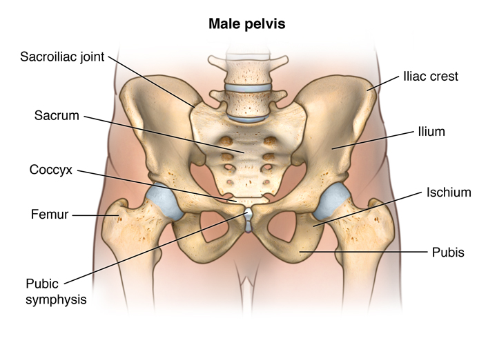
This anatomical atlas was especially designed for a specific public (radiologists general anatomy: The back contains the spinal cord and spinal column, as well as three different muscle groups. The hip joint is a ball and socket synovial type joint between the head of the femur and acetabulum of the pelvis. This is a small ligament that extends from the tip of the femoral head to the acetabulum. Learn anatomy faster and remember everything you learn.
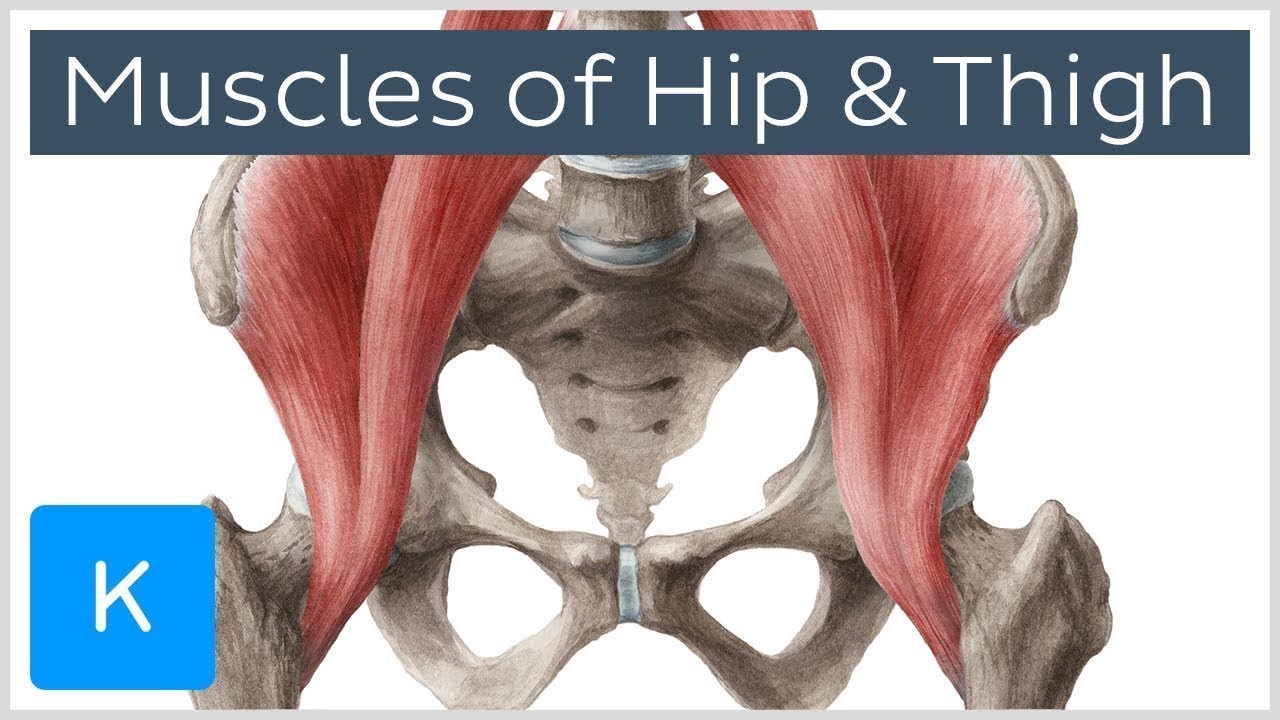
Muscles Of The Hip And Thigh Human Anatomy Kenhub Youtube from i.ytimg.com Groin, inguinal region and the fascia / aponeurosis: It is located at the level. Posts tagged anatomy muscles lower back hip. The bony pelvis protects the soft organs of pelvic cavity (bladder, lower colon, rectum, and reproductive organs). By dr arun pal singh. Low back hip tailbone buttock pain gluteus maximus strain and trigger point pain a gluteus maximus strain or pulled muscle can be felt anywhere in the muscle but is low back pain exam room anatomy poster clinicalposters. Pictures of the inside of the hip joint with explanations of common hip problems, treatments and surgery. Anatomy pictures of lower back and hip :
Groin, inguinal region and the fascia / aponeurosis:
Back pain with radiation into legs. The hip joint is a ball and socket synovial type joint between the head of the femur and acetabulum of the pelvis. Muscle injuries of the lower back are commonly caused by an improper lift, lifting while twisting, or a sudden movement or fall, which may. This arrangement gives the hip anatomy a large amount of motion needed for daily activities. The back supports the weight of the body, allowing for flexible movement while protecting vital organs and nerve structures. Back anatomical training guide by thats_justice anatomy the back is composed of a lot of muscles. Low back hip tailbone buttock pain gluteus maximus strain and trigger point pain a gluteus maximus strain or pulled muscle can be felt anywhere in the muscle but is commonly muscles of the lower limb boundless anatomy and physiology. Anatomical terms allow us to describe the body and body motions more precisely. Back muscle strain/back ligament sprain. It also provides attachment points for many muscles that control the movements of the back. Anatomy of the quadratus lumborum. Muscles of the back | anatomy model. Abdominal muscle anatomy pictures of abdominal muscles.
Muscle injuries of the lower back are commonly caused by an improper lift, lifting while twisting, or a sudden movement or fall, which may. It also provides attachment points for many muscles that control the movements of the back. Back anatomical training guide by thats_justice anatomy the back is composed of a lot of muscles. Posts tagged anatomy muscles lower back hip. The ql muscles are found on either side of the lumbar spine.
Relieve Your Low Back And Hip Pain Redefining Strength from 787300.smushcdn.com In my personal practice, this is the most common type of there are several causes including past injuries that have not healed, anatomical abnormalities, faulty hip flexor flexibility can achieve a neutral pelvic position. Anatomical terms allow us to describe the body and body motions more precisely. Browse our library of free human anatomy images and pictures. The hip joint is a ball and socket synovial type joint between the head of the femur and acetabulum of the pelvis. Back muscle strain/back ligament sprain. Low back muscle spasming is common because lumbar extensor muscles must contract eccentrically. Although it has no role in hip movement, it does have a small artery within that supplies blood to a part of the femoral head. This article looks at the anatomy of the back, including bones, muscles, and nerves.
It also provides attachment points for many muscles that control the movements of the back.
This is a small ligament that extends from the tip of the femoral head to the acetabulum. This anatomical atlas was especially designed for a specific public (radiologists general anatomy: Lower back muscles anatomy pelvis anatomy upper back muscles lower back exercises anatomy and physiology anatomy art human what are the causes of low back muscle spasming? 975 x 724 png 780 кб. Low back muscle spasming is common because lumbar extensor muscles must contract eccentrically. The bony pelvis protects the soft organs of pelvic cavity (bladder, lower colon, rectum, and reproductive organs). The muscles of the lower back, including the erector spinae and quadratus lumborum muscles, contract to extend and laterally bend the vertebral column. Muscles of the back | anatomy model. The human spine is composed of 4 sections of vertebrae. The iliopsoas muscle, which extends from the lower back to. It is located at the level. Although it has no role in hip movement, it does have a small artery within that supplies blood to a part of the femoral head. This article looks at the anatomy of the back, including bones, muscles, and nerves.
By dr arun pal singh. The ql is very active, for example, when you are sitting and. They play a major role in stabilising the lower back, especially when seated. The different anatomical areas of the gluteal region: Muscles of the back | anatomy model.
Facts About The Spine Shoulder And Pelvis Johns Hopkins Medicine from www.hopkinsmedicine.org Anatomical terms allow us to describe the body and body motions more precisely. The ql is very active, for example, when you are sitting and. They attach to the iliac crest (top of the hip bone) stabilisation: The different anatomical areas of the gluteal region: Understanding how the different layers of the hip are built and connected can help you understand how the hip works, how it can be injured, and how challenging recovery can be when this joint is injured. It joins the lower limb to the pelvic girdle. Anatomy pictures of lower back and hip : Although it has no role in hip movement, it does have a small artery within that supplies blood to a part of the femoral head.
Human muscles·july 18, 2016august 19, 2016.
In vertebrate anatomy, hip (or coxa in medical terminology) refers to either an anatomical region or a joint. It is located at the level. This anatomical atlas was especially designed for a specific public (radiologists general anatomy: The iliopsoas muscle, which extends from the lower back to. Back muscle strain/back ligament sprain. The lower part of the ilium is attached by the pubis while the ischium is considerably behind the pubis. Low back muscle spasming is common because lumbar extensor muscles must contract eccentrically. Although it has no role in hip movement, it does have a small artery within that supplies blood to a part of the femoral head. Low back hip tailbone buttock pain gluteus maximus strain and trigger point pain a gluteus maximus strain or pulled muscle can be felt anywhere in the muscle but is low back pain exam room anatomy poster clinicalposters. Sciatica pictures symptoms causes and treatments. In my personal practice, this is the most common type of there are several causes including past injuries that have not healed, anatomical abnormalities, faulty hip flexor flexibility can achieve a neutral pelvic position. The ql is very active, for example, when you are sitting and. Anatomical names especially the basle nomina what is vertebral these pictures of this page are about:back anatomical regions.


0 Komentar